Friday, February 27, 2026

Pleural Sarcoma: Symptoms, Causes, Diagnosis, Treatment, and Prognosis


Pleural Sarcoma: Symptoms, Causes, Diagnosis, Treatment, and Prognosis


Pleural sarcoma is a rare and aggressive type of cancer that develops in the pleura, the thin membrane that lines the lungs and the inside of the chest wall. Unlike more common pleural cancers such as Malignant Pleural Mesothelioma, pleural sarcoma arises from connective tissue cells rather than mesothelial cells. Because of its rarity, pleural sarcoma is often difficult to diagnose, and treatment typically requires a specialized, multidisciplinary approach.

This comprehensive article covers everything you need to know about pleural sarcoma, including its types, causes, symptoms, diagnosis, treatment options, and prognosis.

~What is Pleural Sarcoma?

Pleural sarcoma refers to a group of malignant tumors that originate from the soft tissues of the pleura. The pleura plays an essential role in lung function by producing lubricating fluid that allows smooth movement of the lungs during breathing. When sarcoma develops in this region, it can disrupt breathing and cause significant complications.

Because pleural sarcoma is extremely rare, it is often misdiagnosed as other pleural diseases, including mesothelioma or metastatic cancers. Early recognition and accurate diagnosis are crucial for improving outcomes.

~Types of Pleural Sarcoma

Pleural sarcoma is not a single disease but includes several types of soft tissue sarcomas that may affect the pleura.

1. Synovial Sarcoma of the Pleura

This is one of the most commonly reported pleural sarcomas. Despite its name, it does not originate from synovial tissue but from mesenchymal cells. It may present as a large chest mass.

2. Fibrosarcoma

Fibrosarcoma involves malignant fibroblasts and may develop in the pleura or surrounding chest structures.

3. Leiomyosarcoma

This type arises from smooth muscle cells and may originate from blood vessels in the pleura.

4. Undifferentiated Pleomorphic Sarcoma

Previously known as malignant fibrous histiocytoma, this is an aggressive tumor with no specific differentiation.

~Causes and Risk Factors

The exact cause of pleural sarcoma remains unknown. However, several factors may increase risk.

1. Genetic Mutations

Mutations in tumor suppressor genes and oncogenes can lead to uncontrolled growth of mesenchymal cells.

2. Radiation Exposure

Previous radiation therapy for cancers such as Breast Cancer or Lymphoma may increase the risk of developing secondary sarcomas in the chest.

3. Environmental Factors

Exposure to toxins and industrial chemicals may play a role, although evidence is limited.

Unlike mesothelioma, pleural sarcoma is not strongly linked to asbestos exposure.

~Symptoms of Pleural Sarcoma

Symptoms depend on tumor size, location, and whether the cancer has spread. Because the pleural cavity has space, tumors may grow large before causing symptoms.

Common Symptoms

  • Persistent chest pain

  • Shortness of breath

  • Chronic cough

  • Fatigue

  • Unexplained weight loss

Advanced Symptoms

  • Difficulty breathing

  • Pleural effusion (fluid accumulation)

  • Compression of lung tissue

  • Hoarseness

  • Difficulty swallowing

Pleural effusion can lead to severe breathing problems and often requires drainage.

~How is Pleural Sarcoma Diagnosed?

Diagnosing pleural sarcoma is challenging due to its rarity and similarity to other pleural tumors.

1. Medical History and Physical Examination

Doctors evaluate symptoms, occupational history, and previous radiation exposure.
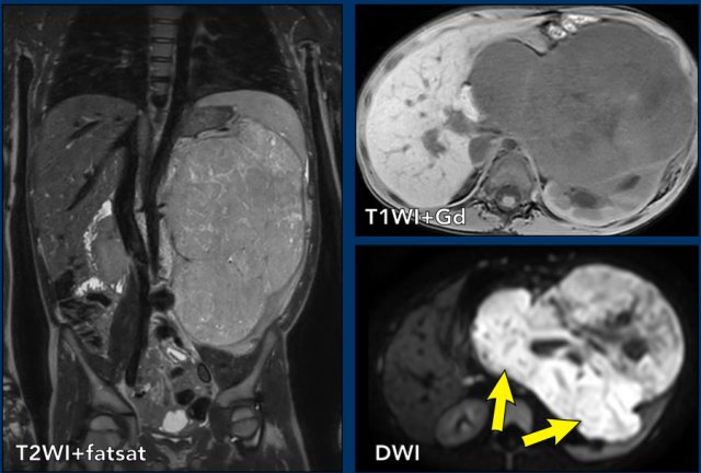
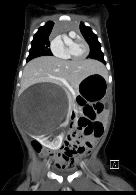
2. Imaging Tests

Key imaging studies include:

  • Chest X-ray

  • CT scan

  • MRI

  • PET scan

These help determine tumor size and spread.

3. Thoracentesis

Fluid from the pleural space may be examined, though it rarely confirms sarcoma.
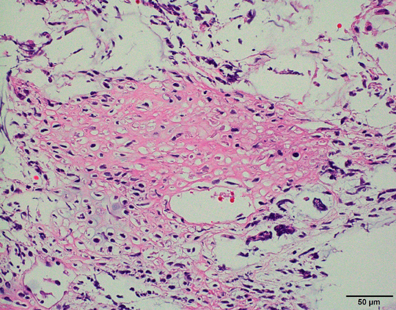
4. Biopsy

A core needle or surgical biopsy is required for definitive diagnosis.

5. Immunohistochemistry and Molecular Testing

These tests help differentiate pleural sarcoma from other cancers such as mesothelioma or metastatic tumors.

~Staging of Pleural Sarcoma

There is no universal staging system specific to pleural sarcoma, but staging is generally based on soft tissue sarcoma guidelines.

Stage I

Localized tumor.

Stage II

Larger tumor or involvement of nearby tissues.

Stage III

Spread to regional lymph nodes.

Stage IV

Metastasis to distant organs such as lungs, liver, or bones.

~Treatment Options for Pleural Sarcoma

Treatment depends on tumor type, stage, and patient health.

1. Surgery

Complete surgical removal is the cornerstone of treatment when possible. Procedures may include:

  • Tumor resection

  • Pleurectomy

  • Removal of affected lung tissue

Surgery offers the best chance for cure in early-stage disease.

2. Chemotherapy

Chemotherapy may be used:

  • Before surgery to shrink tumors

  • After surgery to prevent recurrence

  • In advanced disease

Common drugs include doxorubicin and ifosfamide.

3. Radiation Therapy

Radiation may be used as an adjunct to surgery or for symptom relief.

4. Targeted Therapy

Research is ongoing to identify molecular targets for personalized treatment.

5. Immunotherapy

Some patients may benefit from immune checkpoint inhibitors.

6. Clinical Trials

Due to the rarity of pleural sarcoma, clinical trials are an important option.

~Complications

Pleural sarcoma can lead to several complications:

  • Respiratory failure

  • Tumor compression of lungs

  • Recurrent pleural effusion

  • Metastasis

  • Severe pain

Early intervention helps reduce complications.

~Prognosis and Survival Rate

The prognosis varies depending on several factors.

Factors Affecting Prognosis

  • Tumor type

  • Stage at diagnosis

  • Surgical removal success

  • Patient health

  • Response to therapy

Localized tumors have a better outcome, while advanced disease has a poor prognosis.

~Difference Between Pleural Sarcoma and Mesothelioma

Although both affect the pleura, these cancers differ significantly.

FeaturePleural SarcomaMesothelioma
OriginConnective tissueMesothelial cells
Asbestos linkRareStrong
FrequencyExtremely rareMore common
TreatmentSimilar to soft tissue sarcomaMesothelioma-specific

Accurate diagnosis is essential for selecting the right treatment.

~Prevention and Risk Reduction

Because the exact cause is unclear, prevention focuses on general cancer risk reduction.

Key Measures

  • Avoid unnecessary radiation exposure

  • Limit chemical exposure

  • Maintain regular health checkups

  • Early evaluation of persistent chest symptoms

~Living with Pleural Sarcoma

Living with a rare cancer can be challenging.

Support Strategies

  • Psychological counseling

  • Support groups

  • Nutritional support

  • Pain management

  • Palliative care

These measures improve quality of life.

~Recent Advances in Research

Recent research focuses on:

  • Genetic profiling

  • Targeted therapies

  • Immunotherapy combinations

  • Early detection biomarkers

These developments offer hope for better survival.

~When to See a Doctor

Consult a healthcare provider if you experience:

  • Persistent chest pain

  • Shortness of breath

  • Unexplained weight loss

  • Recurrent pleural fluid

Early diagnosis improves treatment outcomes.

~Conclusion

Pleural sarcoma is a rare but aggressive cancer that requires early detection and specialized care. Because its symptoms mimic other pleural diseases, awareness and timely evaluation are crucial. Advances in surgery, chemotherapy, radiation, and immunotherapy continue to improve outcomes.

If you have persistent chest symptoms or risk factors, seek medical attention promptly. Ongoing research and clinical trials provide hope for better treatments and improved survival in the future.


Hemangiosarcoma: Symptoms, Causes, Diagnosis, Treatment, and Prognosis


Hemangiosarcoma: Symptoms, Causes, Diagnosis, Treatment, and Prognosis


Hemangiosarcoma is a rare and aggressive cancer that develops from the cells lining blood vessels. It belongs to a group of cancers known as soft tissue sarcomas and is characterized by rapid growth and a high potential to spread (metastasize) to other parts of the body. Although it is more commonly seen in animals, particularly dogs, hemangiosarcoma can also occur in humans, where it is considered extremely rare and often challenging to diagnose and treat.

This article provides a comprehensive overview of hemangiosarcoma, including its types, causes, symptoms, diagnosis, treatment options, and prognosis.

~What is Hemangiosarcoma?

Hemangiosarcoma is a malignant tumor that originates in the endothelial cells, which form the inner lining of blood vessels. These tumors can arise in any part of the body where blood vessels are present, but they most commonly affect the skin, liver, spleen, heart, and soft tissues.

Because hemangiosarcoma develops in blood-rich areas, it tends to be highly vascular and prone to bleeding. This characteristic often leads to sudden complications such as internal hemorrhage, which may be life-threatening and sometimes the first sign of the disease.

~Types of Hemangiosarcoma

Hemangiosarcoma can be classified based on the location where it originates. The major types include:

1. Cutaneous Hemangiosarcoma

This form affects the skin and is often associated with chronic sun exposure. It usually appears as a bruise-like lesion or a rapidly growing mass on sun-exposed areas such as the head, neck, or arms.

2. Visceral Hemangiosarcoma

This type develops in internal organs such as the liver, spleen, or heart. It is more aggressive and often diagnosed at an advanced stage due to subtle or absent early symptoms.

3. Cardiac Hemangiosarcoma

When the tumor occurs in the heart, particularly in the right atrium, it may cause fluid accumulation around the heart, leading to symptoms such as shortness of breath and fatigue.

4. Soft Tissue Hemangiosarcoma

This type occurs in muscles and connective tissues and may present as a painless lump that gradually increases in size.

~Causes and Risk Factors

The exact cause of hemangiosarcoma remains unclear. However, researchers have identified several potential risk factors that may increase the likelihood of developing this cancer.

1. Genetic Mutations

Changes in genes that regulate cell growth and blood vessel formation may contribute to the development of hemangiosarcoma.

2. Radiation Exposure

Prior radiation therapy for other cancers has been associated with a higher risk of developing secondary hemangiosarcoma.

3. Chemical Exposure

Exposure to certain industrial chemicals, such as vinyl chloride and arsenic, has been linked to vascular tumors, including hemangiosarcoma.

4. Sun Exposure

Prolonged exposure to ultraviolet (UV) radiation is a known risk factor for cutaneous hemangiosarcoma.

5. Chronic Lymphedema

Long-term swelling due to lymphatic blockage may increase the risk of vascular tumors.

~Symptoms of Hemangiosarcoma

Symptoms vary depending on the location and stage of the tumor. Early stages may not produce noticeable signs, making early diagnosis difficult.

General Symptoms

  • Fatigue

  • Unexplained weight loss

  • Loss of appetite

  • Weakness

  • Fever in some cases

Cutaneous Hemangiosarcoma Symptoms

  • Red, purple, or blue skin lesions

  • Rapidly growing lump

  • Ulceration or bleeding

  • Bruise-like appearance

Visceral Hemangiosarcoma Symptoms

  • Abdominal pain

  • Swelling in the abdomen

  • Internal bleeding

  • Dizziness or fainting

  • Low blood pressure

Cardiac Hemangiosarcoma Symptoms

  • Chest pain

  • Shortness of breath

  • Irregular heartbeat

  • Fluid around the heart

Because these symptoms are non-specific, the condition may initially be mistaken for other diseases.

~How is Hemangiosarcoma Diagnosed?

Diagnosis usually involves a combination of imaging tests, biopsy, and laboratory evaluations.

1. Physical Examination

A thorough clinical examination helps identify suspicious lumps or lesions.

2. Imaging Studies

These include:

  • Ultrasound

  • CT scans

  • MRI

  • PET scans

Imaging helps determine the size, location, and spread of the tumor.

3. Biopsy

A tissue sample is taken and examined under a microscope to confirm the diagnosis.

4. Immunohistochemistry

Special tests are used to identify endothelial markers, which help distinguish hemangiosarcoma from other cancers.

5. Blood Tests

Blood tests may reveal anemia due to bleeding or abnormal liver function.

~Staging of Hemangiosarcoma

Staging helps determine the extent of cancer and guides treatment decisions.

Stage I

Localized tumor without spread.

Stage II

Tumor involving nearby tissues or lymph nodes.

Stage III

Cancer has spread to distant organs such as lungs or liver.

Because hemangiosarcoma spreads quickly, many patients are diagnosed at advanced stages.

~Treatment Options for Hemangiosarcoma

Treatment depends on the location, stage, and overall health of the patient. A multidisciplinary approach is often required.

1. Surgery

Surgical removal of the tumor is the primary treatment when feasible. Complete excision offers the best chance of long-term survival.

However, surgery may not be possible in advanced cases.

2. Chemotherapy

Chemotherapy is commonly used, especially when the cancer has spread. Drugs such as doxorubicin are often part of treatment regimens.

Chemotherapy may help:

  • Slow tumor growth

  • Reduce recurrence

  • Improve survival

3. Radiation Therapy

Radiation may be used:

  • After surgery to eliminate residual cancer cells

  • For palliative care to relieve symptoms

4. Targeted Therapy

Newer therapies focus on specific pathways involved in blood vessel formation.

5. Immunotherapy

Emerging treatments aim to boost the immune system to fight cancer.

6. Clinical Trials

Patients with advanced hemangiosarcoma may benefit from participation in clinical trials exploring novel treatments.

~Complications of Hemangiosarcoma

Due to its aggressive nature, hemangiosarcoma can lead to several complications:

  • Internal bleeding

  • Organ failure

  • Metastasis

  • Anemia

  • Fluid accumulation in organs

Sudden rupture of tumors, especially in the spleen or liver, can cause life-threatening hemorrhage.

~Prognosis and Survival Rate

The prognosis of hemangiosarcoma depends on various factors:

Factors Affecting Prognosis

  • Tumor location

  • Stage at diagnosis

  • Size of the tumor

  • Presence of metastasis

  • Response to treatment

Cutaneous hemangiosarcoma generally has a better prognosis compared to visceral types.

Unfortunately, visceral hemangiosarcoma often has a poor outlook due to late detection and rapid spread.

Early diagnosis and aggressive treatment improve survival rates.

~Prevention and Risk Reduction

There is no guaranteed way to prevent hemangiosarcoma, but certain measures may reduce risk.

Preventive Strategies

  • Limit exposure to harmful chemicals

  • Protect skin from excessive sun exposure

  • Regular health checkups

  • Monitoring high-risk individuals

People with a history of radiation therapy should undergo routine follow-up.

~Living with Hemangiosarcoma

A diagnosis of hemangiosarcoma can be emotionally and physically challenging. Support from healthcare professionals, family, and support groups plays a crucial role in coping.

Lifestyle Tips

  • Maintain a balanced diet

  • Engage in light physical activity

  • Manage stress

  • Follow treatment plans

  • Attend regular follow-up appointments

Palliative care may help improve quality of life in advanced stages.

~Recent Advances in Research

Research in hemangiosarcoma is ongoing, with scientists exploring:

  • Genetic and molecular mechanisms

  • Angiogenesis inhibitors

  • Immune checkpoint therapies

  • Personalized medicine

These advancements offer hope for improved outcomes in the future.

~When to See a Doctor

Seek medical attention if you experience:

  • Persistent unexplained bruising

  • Rapidly growing skin lesions

  • Abdominal pain or swelling

  • Sudden weakness or dizziness

  • Unusual bleeding

Early evaluation increases the chances of timely diagnosis and treatment.

~Conclusion

Hemangiosarcoma is a rare but highly aggressive vascular cancer that requires early detection and prompt treatment. Due to its subtle symptoms and rapid progression, awareness of risk factors and warning signs is essential. Advances in surgical techniques, chemotherapy, targeted therapies, and immunotherapy are improving treatment outcomes and offering hope to patients.

If you notice any suspicious symptoms or skin changes, consult a healthcare professional promptly. Regular checkups and preventive measures can help in early diagnosis and better management of this challenging condition.

Thursday, February 26, 2026

Myxofibrosarcoma: Symptoms, Causes, Diagnosis, Treatment & Survival


Myxofibrosarcoma: Symptoms, Causes, Diagnosis, Treatment & Survival


Myxofibrosarcoma is a rare type of soft tissue sarcoma that typically develops in the connective tissues of the body. It is known for its high recurrence rate and potential to spread if not treated early. This cancer most often affects older adults and usually occurs in the arms and legs. In this detailed article, we will explore the symptoms, causes, diagnosis, treatment options, survival rates, and latest research related to myxofibrosarcoma.

~What Is Myxofibrosarcoma?

Myxofibrosarcoma is a malignant tumor that arises from fibroblasts, the cells responsible for producing connective tissue. It is characterized by a gelatinous (myxoid) matrix and varying degrees of cellularity and aggressiveness.

Previously, myxofibrosarcoma was considered a subtype of Malignant Fibrous Histiocytoma, but advances in pathology and molecular biology have led to its classification as a distinct entity.

This cancer is part of the broader group of Soft Tissue Sarcoma, which includes tumors that develop in muscles, fat, blood vessels, and connective tissues.

~Who Is at Risk?

Myxofibrosarcoma most commonly affects:

  • Adults over the age of 50

  • Slightly more men than women

  • Individuals with a history of radiation exposure

Although rare, it can occur in younger individuals.

~Causes and Risk Factors

The exact cause of myxofibrosarcoma is not well understood. However, several factors may increase the risk:

Genetic Changes

Mutations in certain genes involved in cell growth and repair may lead to tumor formation.

Radiation Exposure

Previous radiation therapy is a known risk factor for soft tissue sarcomas.

Chronic Tissue Injury

Some studies suggest chronic irritation or trauma might contribute, though this is not proven.

Environmental Factors

Research is ongoing to determine the role of environmental toxins.

~Symptoms of Myxofibrosarcoma

Symptoms depend on the tumor’s location and size. In early stages, the tumor may not cause noticeable symptoms.

Common Symptoms

  • A painless lump or swelling under the skin

  • Gradual increase in size

  • Pain or tenderness in later stages

  • Limited movement if near joints

  • Skin ulceration in advanced cases

Because these tumors grow slowly at first, many people ignore early symptoms.

~Where Does Myxofibrosarcoma Occur?

The tumor can develop in various parts of the body, including:

  • Arms and legs (most common)

  • Trunk

  • Head and neck

  • Retroperitoneum (rare)

Lower extremities, especially the thighs, are frequently affected.

~How Is Myxofibrosarcoma Diagnosed?

Early diagnosis is important to prevent spread and recurrence.

Imaging Tests

Doctors use imaging to evaluate the tumor:

  • MRI scan (most effective for soft tissue)

  • CT scan

  • Ultrasound

MRI helps determine tumor extent and involvement of nearby structures.

Biopsy

A biopsy confirms the diagnosis. Tissue samples are examined by a pathologist.

Histological Grading

Tumors are classified based on aggressiveness:

  • Low-grade

  • Intermediate-grade

  • High-grade

High-grade tumors have a higher risk of recurrence and metastasis.

~Staging of Myxofibrosarcoma

Staging determines how far the cancer has spread.

Localized

Cancer is limited to its origin.

Regional

Cancer has spread to nearby tissues.

Metastatic

The tumor spreads to distant organs such as lungs.

Organizations like National Cancer Institute and American Cancer Society provide guidelines for staging and treatment.

~Treatment Options for Myxofibrosarcoma

Treatment depends on tumor size, location, grade, and overall health.

1. Surgery

Surgery is the main treatment. The goal is to remove the tumor with a wide margin to reduce recurrence.

In some cases, reconstructive surgery may be required.

2. Radiation Therapy

Radiation is often used before or after surgery to reduce recurrence risk.

It is particularly helpful when:

  • Tumors are large

  • Surgical margins are close

  • The tumor is in a difficult location

3. Chemotherapy

Chemotherapy is usually recommended for:

  • High-grade tumors

  • Metastatic disease

  • Recurrent tumors

Common drugs include:

  • Doxorubicin

  • Ifosfamide

However, chemotherapy is not always effective in low-grade tumors.

4. Targeted Therapy

Targeted therapy focuses on specific molecular pathways. Research is ongoing to improve outcomes.

5. Immunotherapy

Immunotherapy is being studied in clinical trials for sarcomas.

Research institutions such as St. Jude Children's Research Hospital and MD Anderson Cancer Center are investigating new treatments.

~Recurrence in Myxofibrosarcoma

One of the major challenges is its high recurrence rate.

Why Does It Recur?

  • Microscopic tumor extensions

  • Incomplete surgical removal

  • High-grade tumor biology

Recurrence can occur even years after treatment, so long-term follow-up is essential.

~Survival Rate and Prognosis

Prognosis depends on:

  • Tumor grade

  • Size and depth

  • Surgical margins

  • Patient age

  • Presence of metastasis

Five-Year Survival Rate

  • Low-grade tumors: 70% to 90%

  • High-grade tumors: 40% to 60%

Early detection and complete removal improve survival.

~Complications of Myxofibrosarcoma

Possible complications include:

  • Tumor recurrence

  • Spread to lungs

  • Functional disability

  • Chronic pain

  • Treatment side effects

Regular imaging is needed.

~Can Myxofibrosarcoma Be Prevented?

There is no proven prevention. However:

  • Avoid unnecessary radiation exposure.

  • Regular check-ups for high-risk individuals.

  • Early evaluation of unexplained lumps.

~Living With Myxofibrosarcoma

A cancer diagnosis affects physical and emotional well-being.

Rehabilitation

Physical therapy helps restore movement and strength.

Psychological Support

Counseling and support groups improve coping.

Lifestyle Changes

Healthy diet and regular exercise support recovery.

~Advances in Research

Recent developments include:

  • Genetic profiling

  • Personalized medicine

  • Improved imaging

  • New targeted therapies

Clinical trials offer hope for better treatment options.

~When Should You See a Doctor?

Consult a healthcare professional if you notice:

  • A growing lump

  • Persistent swelling

  • Unexplained pain

  • Changes in skin over a mass

Early evaluation improves outcomes.

~Frequently Asked Questions (FAQs)

Is myxofibrosarcoma curable?

Many patients can be cured if diagnosed early and treated with surgery and radiation.

Is it aggressive?

It can range from slow-growing to highly aggressive depending on grade.

Does it spread?

Yes, especially high-grade tumors may spread to lungs.

Can it return after treatment?

Yes, recurrence is common, requiring long-term monitoring.

~Conclusion

Myxofibrosarcoma is a rare soft tissue cancer with unique challenges, especially its tendency to recur. Early diagnosis, accurate staging, and aggressive treatment significantly improve survival and quality of life. Advances in surgery, radiation, and targeted therapy are helping patients achieve better outcomes.

If you notice unusual swelling or lumps, seek medical advice immediately. Awareness and early intervention remain the key to successful treatment.


Primitive Neuroectodermal Tumor (PNET): Symptoms, Causes, Diagnosis, Treatment & Survival


Primitive Neuroectodermal Tumor (PNET): Symptoms, Causes, Diagnosis, Treatment & Survival


Primitive Neuroectodermal Tumor (PNET) is a rare and aggressive group of cancers that originate from immature nerve cells in the body. These tumors mainly affect children and young adults, although they can occur at any age. Because of their fast-growing nature and ability to spread quickly, early diagnosis and treatment are crucial. This article covers everything you need to know about PNET, including its types, symptoms, causes, treatment options, survival rates, and recent advancements.

~What Is Primitive Neuroectodermal Tumor (PNET)?

Primitive Neuroectodermal Tumor is a type of cancer that develops from primitive (immature) neuroectodermal cells. These cells are responsible for forming the nervous system during early development. When these cells grow abnormally, they form tumors that can occur in the brain, spinal cord, or other parts of the body.

PNET belongs to the family of small round blue cell tumors and is closely related to the Ewing Sarcoma group. Some experts classify certain PNETs as part of the Ewing sarcoma spectrum due to similar genetic and biological characteristics.

~Types of Primitive Neuroectodermal Tumors

PNETs are classified based on where they develop in the body. The main types include:

1. Central Nervous System (CNS) PNET

These tumors occur in the brain or spinal cord. They are highly aggressive and may spread through cerebrospinal fluid.

Common subtypes include:

  • Medulloblastoma (most common in children)

  • Pineoblastoma

  • Other embryonal tumors

2. Peripheral PNET (pPNET)

Peripheral PNETs develop outside the brain and spinal cord. They commonly affect the:

  • Chest wall

  • Abdomen

  • Pelvis

  • Limbs

Peripheral PNET is closely related to Askin Tumor when it occurs in the chest.

~Who Is at Risk?

PNETs primarily affect:

  • Children and adolescents

  • Young adults

  • Slightly more males than females

Although the exact cause is unknown, some genetic and environmental factors may increase risk.

~Causes and Risk Factors of PNET

The precise cause of Primitive Neuroectodermal Tumors remains unclear. However, researchers have identified several contributing factors:

Genetic Mutations

Many PNETs are linked to chromosomal abnormalities. One common mutation is the translocation between chromosomes 11 and 22, which also occurs in Ewing sarcoma.

Family History

Most cases are not inherited, but rare genetic syndromes may increase risk.

Environmental Factors

Although no clear environmental cause has been proven, ongoing research is examining possible links.

~Symptoms of Primitive Neuroectodermal Tumor

Symptoms vary depending on the tumor’s location and size. Because these tumors grow quickly, symptoms may develop rapidly.

Symptoms of CNS PNET

  • Persistent headaches

  • Nausea and vomiting

  • Vision problems

  • Balance issues

  • Seizures

  • Weakness or numbness

Symptoms of Peripheral PNET

  • Pain or swelling in affected areas

  • Chest pain or breathing difficulty

  • Abdominal discomfort

  • Fatigue

  • Weight loss

In many cases, symptoms appear suddenly and worsen quickly.

~How Is PNET Diagnosed?

Early diagnosis improves outcomes. Doctors use a combination of imaging, biopsy, and laboratory tests.

Imaging Tests

These help detect tumor location and spread:

  • MRI scan

  • CT scan

  • PET scan

MRI is especially useful for brain and spinal tumors.

Biopsy

A biopsy confirms diagnosis by examining tumor cells under a microscope.

Molecular and Genetic Testing

Genetic testing helps identify specific mutations and guide treatment.

Lumbar Puncture

For CNS tumors, cerebrospinal fluid may be examined to check for spread.

Organizations such as American Cancer Society and National Cancer Institute recommend multidisciplinary evaluation for rare tumors like PNET.

~Staging of Primitive Neuroectodermal Tumor

Staging helps determine tumor spread and guides treatment.

Localized

The tumor is confined to its original site.

Regional

Cancer has spread to nearby tissues or lymph nodes.

Metastatic

The tumor has spread to distant organs such as lungs, bones, or brain.

PNET is often aggressive and may be metastatic at diagnosis.

~Treatment Options for Primitive Neuroectodermal Tumor

Treatment depends on tumor type, location, stage, and patient age. A combination of therapies is usually required.

1. Surgery

Surgical removal of the tumor is often the first step. The goal is to remove as much cancer as possible.

For brain tumors, surgery may be complex due to sensitive areas.

2. Chemotherapy

Chemotherapy is a key treatment for PNET. It targets fast-growing cancer cells and is often used before or after surgery.

Common drugs include:

  • Vincristine

  • Cyclophosphamide

  • Doxorubicin

  • Ifosfamide

Combination chemotherapy improves survival.

3. Radiation Therapy

Radiation is used to destroy remaining cancer cells after surgery.

For CNS PNET:

  • Craniospinal radiation may be required.

However, doctors carefully consider long-term side effects, especially in children.

4. Targeted Therapy

New treatments target specific genetic mutations. These therapies are more precise and may cause fewer side effects.

Research is ongoing in this field.

5. Immunotherapy

Immunotherapy boosts the immune system to fight cancer. Although still experimental for PNET, clinical trials are promising.

6. Stem Cell Transplant

High-dose chemotherapy followed by stem cell transplant may be recommended for high-risk or recurrent cases.

~Side Effects of PNET Treatment

Side effects vary depending on treatment:

Short-Term Effects

  • Nausea

  • Hair loss

  • Fatigue

  • Infection risk

Long-Term Effects

  • Growth and development issues

  • Learning difficulties

  • Hormonal imbalance

  • Secondary cancers

Long-term follow-up care is essential.

~Survival Rate and Prognosis

Survival depends on several factors:

  • Tumor location

  • Stage at diagnosis

  • Age

  • Response to treatment

CNS PNET Survival

Five-year survival ranges between 50% and 70% for children, depending on risk factors.

Peripheral PNET Survival

With aggressive treatment, survival may reach 60% to 75%.

Early diagnosis significantly improves outcomes.

~Complications of Primitive Neuroectodermal Tumor

Possible complications include:

  • Tumor recurrence

  • Metastasis

  • Neurological deficits

  • Treatment-related complications

Regular monitoring is necessary.

~Can PNET Be Prevented?

There is currently no proven way to prevent Primitive Neuroectodermal Tumor. However:

  • Early detection improves outcomes.

  • Genetic counseling may help high-risk families.

  • Awareness of symptoms is crucial.

~Living With Primitive Neuroectodermal Tumor

A PNET diagnosis can be overwhelming. Support from healthcare teams, family, and support groups is important.

Emotional and Psychological Support

Patients and families benefit from counseling and support groups.

Rehabilitation

Physical and occupational therapy help recovery.

Education Support

Children may need special educational assistance after treatment.

~Advances in PNET Research

Medical research is improving diagnosis and treatment.

Precision Medicine

Genetic profiling helps tailor treatments.

Clinical Trials

New drugs and therapies are under investigation.

Organizations like St. Jude Children's Research Hospital play a major role in advancing pediatric cancer treatment.

~When Should You See a Doctor?

Consult a doctor if you notice:

  • Persistent headaches

  • Unexplained swelling or pain

  • Neurological symptoms

  • Rapid weight loss

Early evaluation saves lives.

~Frequently Asked Questions (FAQs)

Is Primitive Neuroectodermal Tumor curable?

Some patients can be cured, especially when diagnosed early and treated aggressively.

Is PNET hereditary?

Most cases are not inherited, although genetic factors may play a role.

Can adults develop PNET?

Yes, but it is more common in children and young adults.

How fast does PNET grow?

These tumors grow rapidly and require immediate treatment.

~Conclusion

Primitive Neuroectodermal Tumor (PNET) is a rare but aggressive cancer that requires early diagnosis and multidisciplinary treatment. Advances in surgery, chemotherapy, radiation, and targeted therapies are improving survival rates. Continued research offers hope for better treatments and outcomes in the future.

If you or a loved one experience symptoms, seek medical care promptly. Awareness, early detection, and modern treatment options can make a significant difference in survival and quality of life.


Wednesday, February 25, 2026

Langerhans Cell Histiocytosis: Symptoms, Causes, Diagnosis, Treatment, and Survival


Langerhans Cell Histiocytosis: Symptoms, Causes, Diagnosis, Treatment, and Survival 

~Introduction to Langerhans Cell Histiocytosis


Langerhans Cell Histiocytosis (LCH)
is a rare disorder characterized by the abnormal accumulation of Langerhans cells, a type of immune cell that normally helps protect the body against infections. In this disease, these cells grow uncontrollably and can form tumors or lesions in different parts of the body.

LCH can affect both children and adults, although it is more commonly diagnosed in young children, especially those under the age of 10. The disease ranges from mild, single-organ involvement to severe, life-threatening multisystem disease.

This guide provides detailed information about Langerhans Cell Histiocytosis, including symptoms, causes, risk factors, diagnosis, treatment, prognosis, and long-term outcomes.

~What Is Langerhans Cell Histiocytosis?

LCH is considered a disorder of the immune system and is sometimes classified as a rare cancer-like condition. It occurs when Langerhans cells multiply excessively and infiltrate tissues, leading to inflammation and damage.

These abnormal cells can affect:

  • Bones

  • Skin

  • Lymph nodes

  • Lungs

  • Liver

  • Spleen

  • Bone marrow

  • Brain and central nervous system

The severity of LCH varies greatly depending on the organs involved and the extent of the disease.

~How Common Is Langerhans Cell Histiocytosis?

LCH is a rare condition:

  • Occurs in approximately 2 to 5 children per million each year

  • More common in children than adults

  • Slightly more common in boys

  • Adult cases are less frequent but increasingly recognized

Because it is rare, diagnosis is sometimes delayed or mistaken for other conditions.

~Types of Langerhans Cell Histiocytosis

LCH is categorized based on how many organs are involved:

1. Single-System LCH

Only one organ or body system is affected. Common forms include:

  • Bone lesions

  • Skin involvement

  • Lymph node disease

This type generally has a good prognosis.

2. Multisystem LCH

Multiple organs are affected simultaneously. It may involve:

  • Liver

  • Spleen

  • Bone marrow

This form can be serious and requires aggressive treatment.

3. High-Risk LCH

High-risk organs include:

  • Liver

  • Spleen

  • Bone marrow

Involvement of these organs is associated with poorer outcomes.

~Causes of Langerhans Cell Histiocytosis

The exact cause of LCH is still not fully understood. However, recent research suggests it is linked to genetic mutations in immune cells.

Genetic Mutations

Many patients with LCH have mutations in the BRAF gene, especially the BRAF V600E mutation. Other gene alterations may include:

  • MAP2K1

  • ARAF

These mutations cause uncontrolled growth and survival of Langerhans cells.

~Is LCH a Cancer?

LCH is not always classified as a traditional cancer. However, because it involves abnormal cell proliferation and may require chemotherapy, it is often treated similarly to cancer.

Some experts now consider LCH part of a group of disorders called histiocytic neoplasms.

~Risk Factors

Although the exact cause is unknown, certain factors may increase the risk:

  • Young age

  • Male gender

  • Family history (rare)

  • Environmental exposures (not proven)

Smoking is strongly associated with pulmonary LCH in adults.

~Symptoms of Langerhans Cell Histiocytosis

Symptoms depend on the organs involved.

Bone Symptoms

Bone involvement is the most common form.

  • Bone pain

  • Swelling

  • Tenderness

  • Fractures

  • Skull lesions

  • Jaw problems

Skin Symptoms

  • Rashes resembling eczema

  • Scaly or crusted skin

  • Lesions on scalp or trunk

  • Persistent diaper rash in infants

Lymph Node Symptoms

  • Enlarged lymph nodes

  • Swelling in neck, armpits, or groin

Lung Symptoms (Pulmonary LCH)

  • Chronic cough

  • Shortness of breath

  • Chest pain

  • Pneumothorax (collapsed lung)

Hormonal and Brain Symptoms

When the brain or pituitary gland is involved:

  • Excessive thirst and urination (diabetes insipidus)

  • Growth problems

  • Puberty delays

  • Hormonal imbalances

Systemic Symptoms

  • Fever

  • Fatigue

  • Weight loss

  • Anemia

~Complications of LCH

If untreated, LCH can lead to:

  • Organ failure

  • Lung damage

  • Bone deformities

  • Neurological problems

  • Permanent hormone deficiencies

Early diagnosis improves outcomes.

~How Is Langerhans Cell Histiocytosis Diagnosed?

1. Physical Examination

Doctors evaluate symptoms, skin lesions, and bone tenderness.

2. Imaging Tests

  • X-rays for bone lesions

  • CT scans for lung involvement

  • MRI for brain or spinal disease

  • PET scans to detect active disease

3. Biopsy

A biopsy is required to confirm diagnosis. Tissue samples are examined for:

  • Langerhans cells

  • Specific markers like CD1a and langerin

4. Blood and Bone Marrow Tests

Used to assess organ function and detect multisystem disease.

~Staging and Risk Assessment

Patients are classified based on:

  • Number of organs involved

  • Presence of high-risk organs

  • Response to initial therapy

This helps guide treatment and predict prognosis.

~Treatment of Langerhans Cell Histiocytosis

Treatment varies depending on disease severity.

1. Observation

Some mild cases, especially single bone lesions, may resolve without aggressive treatment.

2. Surgery

Surgery may be used to:

  • Remove isolated bone lesions

  • Confirm diagnosis

3. Corticosteroids

Steroids reduce inflammation and immune activity.

They may be given:

  • Locally

  • Systemically

4. Chemotherapy

For multisystem disease or high-risk cases.

Common drugs include:

  • Vinblastine

  • Prednisone

  • Methotrexate

  • Cytarabine

Chemotherapy improves survival in severe cases.

5. Targeted Therapy

Patients with BRAF mutations may benefit from targeted drugs such as:

  • BRAF inhibitors

  • MEK inhibitors

These treatments represent a major breakthrough.

6. Radiation Therapy

Rarely used but may help in:

  • Painful bone lesions

  • Localized disease

7. Lung Treatment

For pulmonary LCH:

  • Smoking cessation is essential

  • Oxygen therapy

  • Lung transplant in severe cases

~Side Effects of Treatment

Short-Term Effects

  • Nausea

  • Fatigue

  • Hair loss

  • Increased infection risk

Long-Term Effects

  • Hormonal deficiencies

  • Fertility issues

  • Learning difficulties

  • Growth problems

Regular monitoring is required.

~Prognosis and Survival Rates

Prognosis depends on age, organs involved, and response to therapy.

Survival Rates

  • Single-system LCH: Excellent prognosis

  • Multisystem without high-risk organs: Good survival

  • High-risk LCH: Survival rates range from 60% to 80%

Early treatment significantly improves outcomes.

~Recurrence of LCH

Relapse is common, especially in:

  • Multisystem disease

  • BRAF mutation-positive cases

Patients require long-term follow-up.

~Langerhans Cell Histiocytosis in Adults

Adult LCH differs in several ways:

  • More lung involvement

  • Strong association with smoking

  • Chronic course

  • Hormonal complications

Management may vary compared to children.

~Living with LCH

Long-term care focuses on:

  • Monitoring relapse

  • Hormone replacement therapy

  • Physical rehabilitation

  • Psychological support

Many patients lead normal lives with appropriate treatment.

~Can LCH Be Prevented?

Currently, there is no known method to prevent LCH.

However:

  • Avoiding smoking reduces pulmonary risk.

  • Early medical attention for persistent symptoms is crucial.

~Latest Research and Advances

New developments in LCH include:

  • Molecular-targeted therapies

  • Precision medicine

  • Improved genetic testing

  • Better risk classification

  • Reduced chemotherapy toxicity

These advances are improving survival and quality of life.

~Frequently Asked Questions (FAQs)

Is Langerhans Cell Histiocytosis life-threatening?

It can be, especially in multisystem high-risk disease. However, many patients recover.

Is LCH hereditary?

Most cases are not inherited.

Can LCH come back?

Yes, relapse is possible.

Is LCH curable?

Many cases, especially single-system disease, are curable.

~Conclusion

Langerhans Cell Histiocytosis is a rare but complex condition that ranges from mild, localized disease to severe multisystem involvement. Advances in genetic research, targeted therapy, and personalized treatment have dramatically improved patient outcomes.

Early diagnosis, multidisciplinary care, and long-term monitoring are essential for managing LCH effectively. With ongoing research and improved treatment strategies, the future outlook for patients with LCH continues to improve.


Medulloblastoma: Symptoms, Causes, Diagnosis, Treatment, and Survival Rates


Medulloblastoma: Symptoms, Causes, Diagnosis, Treatment, and Survival Rates 

~Introduction to Medulloblastoma


Medulloblastoma
is a fast-growing, malignant brain tumor that primarily develops in the cerebellum—the lower back part of the brain responsible for coordination, balance, and motor control. It is classified as a primitive neuroectodermal tumor (PNET) and is considered one of the most common malignant brain tumors in children.

Although medulloblastoma predominantly affects children between the ages of 3 and 8, it can also occur in adolescents and, rarely, adults. Early diagnosis and advances in multimodal therapy—including surgery, radiation, and chemotherapy—have significantly improved survival rates over the past few decades.

This comprehensive guide provides an in-depth overview of medulloblastoma, covering key aspects such as symptoms, causes, risk factors, molecular subtypes, diagnosis, treatment options, prognosis, survival rates, and long-term outcomes.

~What Is Medulloblastoma?

Medulloblastoma is a Grade 4 tumor according to the World Health Organization (WHO) classification. Grade 4 means the tumor is highly aggressive, grows rapidly, and has the potential to spread (metastasize) to other parts of the brain and spinal cord via cerebrospinal fluid (CSF).

Unlike many adult brain tumors, medulloblastoma often spreads along the spinal cord at the time of diagnosis, making comprehensive evaluation essential.

~How Common Is Medulloblastoma?

  • Accounts for approximately 20% of all childhood brain tumors

  • Most common malignant brain tumor in children

  • Rare in adults (less than 1% of adult brain tumors)

  • Slightly more common in males than females

In India and globally, improved neuroimaging and pediatric oncology services have enhanced detection and management outcomes.

~Types and Molecular Subgroups of Medulloblastoma

Modern research has identified four major molecular subgroups, each with distinct genetic characteristics and outcomes:

1. WNT Subgroup

  • Best prognosis

  • Often occurs in older children

  • Survival rate exceeds 90% with treatment

2. SHH (Sonic Hedgehog) Subgroup

  • Common in infants and adults

  • Intermediate prognosis

  • Associated with genetic mutations

3. Group 3

  • Most aggressive form

  • Higher risk of metastasis

  • Lower survival rate

4. Group 4

  • Most common subtype

  • Intermediate prognosis

  • Frequently diagnosed in school-age children

Understanding these subgroups helps doctors tailor treatment and predict outcomes more accurately.

~Causes of Medulloblastoma

The exact cause of medulloblastoma remains unknown. However, it is believed to result from genetic mutations that cause uncontrolled cell growth in cerebellar cells.

Genetic Factors

Certain inherited conditions increase risk:

  • Gorlin syndrome

  • Turcot syndrome

  • Li-Fraumeni syndrome

  • Familial adenomatous polyposis (FAP)

Most cases, however, are sporadic, meaning they occur without a clear inherited cause.

~Risk Factors

While medulloblastoma has no well-defined environmental causes, known risk factors include:

  • Young age (most common in children under 10)

  • Male gender

  • Genetic predisposition syndromes

  • Prior radiation exposure (rare)

~Symptoms of Medulloblastoma

Symptoms usually develop due to increased intracranial pressure or cerebellar dysfunction.

Early Symptoms

  • Persistent morning headaches

  • Nausea and vomiting

  • Dizziness

  • Fatigue

  • Irritability in young children

Neurological Symptoms

  • Poor balance (ataxia)

  • Difficulty walking

  • Clumsiness

  • Double vision

  • Abnormal eye movements

  • Slurred speech

Advanced Symptoms

  • Back pain (if spread to spinal cord)

  • Weakness in limbs

  • Behavioral changes

  • Enlarged head in infants

Symptoms may worsen rapidly because medulloblastoma grows quickly.

~How Is Medulloblastoma Diagnosed?

Early diagnosis is critical for improving survival outcomes.

1. Neurological Examination

Doctors assess coordination, reflexes, vision, and motor function.

2. MRI Scan

MRI of the brain and spine is the gold standard imaging method. It shows tumor location, size, and possible spread.

3. CT Scan

May be used in emergency situations.

4. Lumbar Puncture (Spinal Tap)

Checks cerebrospinal fluid (CSF) for cancer cells.

5. Biopsy and Surgical Resection

Tumor tissue is examined under a microscope to confirm diagnosis and determine molecular subtype.

~Staging of Medulloblastoma

Staging determines whether the tumor has spread:

  • M0 – No metastasis

  • M1 – Cancer cells in CSF

  • M2 – Spread within brain

  • M3 – Spread to spinal cord

  • M4 – Spread outside central nervous system (rare)

Risk classification is generally divided into:

  • Standard-risk

  • High-risk

~Treatment of Medulloblastoma

Treatment typically involves a combination of therapies:

1. Surgery

The first step is maximal safe surgical removal of the tumor.

Goals:

  • Remove as much tumor as possible

  • Relieve pressure in the brain

  • Obtain tissue for diagnosis

Complete resection improves survival chances.

2. Radiation Therapy

Radiation is commonly used after surgery, especially in children over 3 years old.

Types include:

  • Craniospinal irradiation (CSI)

  • Boost radiation to tumor bed

Radiation helps eliminate remaining cancer cells.

3. Chemotherapy

Chemotherapy is used:

  • After surgery

  • Along with radiation

  • In infants to delay radiation

Common drugs include:

  • Cisplatin

  • Vincristine

  • Cyclophosphamide

  • Carboplatin

Chemotherapy improves long-term survival but may have side effects.

4. Targeted Therapy

In SHH subgroup tumors, targeted drugs that block specific molecular pathways are being studied.

5. Clinical Trials

Many patients benefit from enrollment in clinical trials exploring:

  • Reduced radiation doses

  • Immunotherapy

  • Precision medicine approaches

~Side Effects of Treatment

Because treatment affects the developing brain, long-term effects are common:

Short-Term Effects

  • Hair loss

  • Fatigue

  • Nausea

  • Increased infection risk

Long-Term Effects

  • Learning difficulties

  • Memory problems

  • Hormonal imbalance

  • Hearing loss

  • Growth issues

  • Emotional challenges

Regular follow-up care is essential.

~Survival Rates for Medulloblastoma

Survival depends on age, molecular subtype, and spread.

5-Year Survival Rates:

  • Standard-risk children: 70%–85%

  • WNT subgroup: Over 90%

  • High-risk cases: 50%–70%

  • Adults: 60%–80%

Advancements in treatment have significantly improved outcomes compared to previous decades.

~Prognosis Factors

Better prognosis is associated with:

  • Complete tumor removal

  • No metastasis

  • WNT molecular subtype

  • Older age at diagnosis

  • Early detection

Poor prognosis factors include:

  • Group 3 subtype

  • Metastatic disease

  • Residual tumor after surgery

~Can Medulloblastoma Be Prevented?

There is currently no known way to prevent medulloblastoma. However:

  • Genetic counseling may help families with inherited syndromes.

  • Early evaluation of persistent neurological symptoms is crucial.

~Medulloblastoma in Adults

Although rare, adult medulloblastoma does occur.

Differences include:

  • More common SHH subtype

  • Better tolerance to radiation

  • Slightly different chemotherapy regimens

Treatment principles remain similar to pediatric cases.

~Recurrence of Medulloblastoma

Unfortunately, recurrence can occur, especially in high-risk cases.

Common recurrence locations:

  • Tumor bed

  • Spinal cord

  • Brain lining

Treatment options for recurrence may include:

  • Additional chemotherapy

  • Re-irradiation

  • Clinical trials

  • Stem cell transplant (in select cases)

~Living After Medulloblastoma

Survivorship care focuses on:

  • Neurocognitive rehabilitation

  • Physical therapy

  • Endocrine management

  • Psychological support

  • Educational assistance

Multidisciplinary follow-up care is essential for improving quality of life.

~Frequently Asked Questions (FAQs)

Is medulloblastoma curable?

Yes, many children with standard-risk medulloblastoma can be cured with modern treatment.

Is medulloblastoma hereditary?

Most cases are not inherited, but some genetic syndromes increase risk.

How fast does medulloblastoma grow?

It is a fast-growing, aggressive tumor that requires urgent treatment.

Can adults get medulloblastoma?

Yes, but it is rare.

~Latest Research and Future Directions

Emerging research focuses on:

  • Molecular-targeted therapies

  • Reduced radiation protocols

  • Immunotherapy

  • Genetic profiling

  • Personalized treatment plans

The goal is to improve survival while reducing long-term side effects.

~Conclusion

Medulloblastoma is a serious and aggressive brain tumor primarily affecting children, but significant advances in surgery, radiation therapy, chemotherapy, and molecular research have dramatically improved survival outcomes.

Early recognition of symptoms such as persistent headaches, balance problems, and vomiting is critical. Multimodal treatment strategies tailored to molecular subtype now offer hope for higher survival rates and better quality of life.

Ongoing research continues to refine therapies and reduce treatment-related complications, making the future increasingly promising for patients diagnosed with medulloblastoma.


Rhabdoid Tumor: Causes, Symptoms, Diagnosis, Treatment & Prognosis


Rhabdoid Tumor: Causes, Symptoms, Diagnosis, Treatment & Prognosis

~Introduction

Rhabdoid tumor is a rare but highly aggressive form of cancer that primarily affects infants and very young children. First identified in the kidney, this tumor is now known to occur in multiple parts of the body, including the brain and soft tissues. Due to its rapid growth and early spread, rhabdoid tumor requires immediate medical attention and specialized treatment.

This detailed article explains everything you need to know about rhabdoid tumor — including its types, causes, symptoms, diagnosis, treatment options, survival rates, and ongoing research developments.

~What is a Rhabdoid Tumor?

A rhabdoid tumor is a fast-growing cancer made up of abnormal cells that appear “rhabdoid” (resembling muscle cells) under a microscope. Despite the name, these tumors do not arise from muscle tissue. Instead, they are linked to genetic mutations affecting tumor suppressor genes.

Rhabdoid tumors most commonly affect:

  • The kidneys

  • The brain

  • Soft tissues throughout the body

These cancers are most frequently diagnosed in children under the age of 3, although rare cases can occur in older children and adults.

~Types of Rhabdoid Tumors

1. Malignant Rhabdoid Tumor of the Kidney (MRTK)

Image

Image

Image

Image

Malignant Rhabdoid Tumor of the Kidney (MRTK) was initially thought to be a variant of Wilms tumor but is now recognized as a distinct and more aggressive cancer. It primarily affects infants and toddlers.

It often presents as a large abdominal mass and may spread rapidly to the lungs or brain.

2. Atypical Teratoid/Rhabdoid Tumor (AT/RT)

Image

Image

Image

Image

Atypical Teratoid/Rhabdoid Tumor (AT/RT) is a rhabdoid tumor that occurs in the central nervous system, most commonly in the cerebellum or brainstem. It is one of the most aggressive pediatric brain tumors.

Symptoms depend on tumor location and may include headaches, vomiting, or developmental delays.

3. Extrarenal Rhabdoid Tumor

These tumors develop outside the kidney and brain, often in soft tissues such as:

  • Neck

  • Liver

  • Chest

  • Pelvis

They are rare but equally aggressive.

~Causes and Genetic Factors

Rhabdoid tumors are strongly associated with mutations in the SMARCB1 gene, also known as the INI1 gene. This gene normally helps suppress tumor growth. When it is missing or mutated, uncontrolled cell division occurs.

Some children inherit this mutation, leading to a condition called Rhabdoid Tumor Predisposition Syndrome (RTPS). However, most cases occur sporadically without family history.

Key genetic aspects:

  • Loss of SMARCB1 gene function

  • Rarely, mutation in SMARCA4 gene

  • May be inherited or spontaneous

Genetic counseling is recommended for affected families.

~Risk Factors

Although rhabdoid tumors are rare, certain factors increase risk:

  • Age under 3 years

  • Genetic mutation in SMARCB1

  • Family history of rhabdoid tumor

  • Rhabdoid Tumor Predisposition Syndrome

There are no known environmental or lifestyle causes.

~Symptoms of Rhabdoid Tumor

Symptoms vary depending on tumor location.

Kidney Tumor Symptoms:

  • Abdominal swelling or mass

  • Blood in urine

  • Fever

  • Irritability

  • Weight loss

Brain Tumor (AT/RT) Symptoms:

  • Persistent headache

  • Vomiting

  • Balance problems

  • Seizures

  • Enlarged head size in infants

Soft Tissue Tumor Symptoms:

  • Swelling or lump

  • Pain

  • Breathing difficulty (if chest involved)

Because these tumors grow rapidly, symptoms often worsen quickly.

~Diagnosis of Rhabdoid Tumor

Early diagnosis is critical due to the tumor’s aggressive nature.

1. Physical Examination

Doctors check for lumps, neurological changes, or abdominal enlargement.

2. Imaging Tests

  • Ultrasound

  • CT scan

  • MRI

MRI is especially important for brain tumors.

3. Biopsy

A tissue sample confirms the diagnosis. Pathologists look for:

  • Rhabdoid cell appearance

  • Loss of INI1 protein (key diagnostic marker)

4. Genetic Testing

Testing for SMARCB1 mutation helps confirm diagnosis and guide family counseling.

~Staging and Spread

Rhabdoid tumors are often advanced at diagnosis. They can spread (metastasize) to:

  • Lungs

  • Brain

  • Liver

  • Lymph nodes

Staging depends on:

  • Tumor size

  • Spread to distant organs

  • Lymph node involvement

Because of early metastasis, aggressive treatment is required.

~Treatment Options

Treatment is intensive and usually combines multiple therapies.

1. Surgery

Complete surgical removal of the tumor is the primary goal when possible.

  • Kidney removal (nephrectomy) for MRTK

  • Brain tumor resection for AT/RT

However, complete removal is not always achievable.

2. Chemotherapy

High-dose chemotherapy is standard treatment.

Common drugs include:

  • Vincristine

  • Cyclophosphamide

  • Doxorubicin

  • Cisplatin

  • Etoposide

Chemotherapy may be given before or after surgery.

3. Radiation Therapy

Used particularly in brain tumors, especially in older children. Radiation may be limited in infants due to long-term side effects.

4. Stem Cell Transplant

In some cases, high-dose chemotherapy followed by stem cell rescue is used to improve survival chances.

5. Targeted Therapy and Clinical Trials

Researchers are studying new treatments targeting genetic mutations. Immunotherapy and molecular-targeted drugs are under investigation.

Participation in clinical trials is often recommended.

~Prognosis and Survival Rate

Rhabdoid tumors are among the most aggressive childhood cancers.

Overall survival rates remain low, especially in:

  • Infants under 1 year

  • Metastatic disease

  • Incomplete tumor removal

However, survival has improved with aggressive multimodal therapy.

Early-stage diagnosis and complete surgical removal significantly improve outcomes.

~Complications

Possible complications include:

  • Tumor recurrence

  • Treatment-related side effects

  • Organ damage

  • Developmental delays (brain tumor cases)

Long-term follow-up is essential.

~Follow-Up and Survivorship

Children who survive require ongoing monitoring for:

  • Recurrence

  • Growth and development issues

  • Secondary cancers

  • Cognitive function (in brain tumor survivors)

Rehabilitation, educational support, and psychological counseling may be needed.

~Advances in Research

Recent research focuses on:

  • Targeting SMARCB1 mutation pathways

  • Epigenetic therapies

  • Precision medicine approaches

  • Improved chemotherapy protocols

Genomic studies are helping develop more personalized treatments.

~Emotional and Family Support

A diagnosis of rhabdoid tumor is devastating for families. Support resources include:

  • Pediatric oncology teams

  • Genetic counselors

  • Support groups

  • Palliative care specialists

Emotional and psychological care is crucial alongside medical treatment.

~Prevention and Screening

There is no known way to prevent rhabdoid tumors.

However, children with known SMARCB1 mutations may undergo:

  • Regular MRI scans

  • Routine physical exams

Early detection may improve survival.

~Frequently Asked Questions (FAQs)

1. Is rhabdoid tumor hereditary?
Most cases are not inherited, but some are linked to genetic mutations passed down in families.

2. What age is most affected?
Children under 3 years are most commonly diagnosed.

3. Is rhabdoid tumor curable?
It can be treated, but it is aggressive and survival depends on early diagnosis and response to therapy.

4. How rare is rhabdoid tumor?
It is extremely rare, accounting for a small percentage of pediatric cancers.

5. Can adults get rhabdoid tumors?
Rarely, but most cases occur in infants and toddlers.

~Conclusion

Rhabdoid tumor is a rare and highly aggressive childhood cancer that requires urgent, specialized treatment. Whether occurring in the kidney, brain, or soft tissues, it is linked to genetic mutations and tends to spread quickly.

Although prognosis remains challenging, advances in surgery, chemotherapy, stem cell therapy, and genetic research are improving outcomes. Early detection, comprehensive treatment, and strong family support play vital roles in managing this condition.


Hepatoblastoma: Causes, Symptoms, Diagnosis, Treatment, and Survival


Hepatoblastoma: Causes, Symptoms, Diagnosis, Treatment, and Survival

~Introduction


Hepatoblastoma is a rare but serious type of liver cancer that primarily affects infants and young children. Although uncommon, it is the most frequent malignant liver tumor in pediatric patients. With advances in medical science, early diagnosis and modern treatment approaches have significantly improved survival rates. However, awareness among parents, caregivers, and healthcare providers remains essential to ensure timely intervention.

This comprehensive guide explores hepatoblastoma in detail, including its causes, symptoms, risk factors, stages, diagnosis, treatment options, and long-term outlook. Whether you are a parent, student, or healthcare professional, this article provides reliable information to help you understand this condition.

~What is Hepatoblastoma?

Hepatoblastoma is a cancer that develops in the liver, most commonly in children under the age of 3. The tumor originates from immature liver cells called hepatoblasts, which normally develop into mature liver tissue. When these cells grow abnormally and uncontrollably, they form a tumor.

Unlike adult liver cancers, hepatoblastoma is not typically associated with alcohol use or chronic liver disease. Instead, it is considered a pediatric embryonal tumor, meaning it develops during early childhood.

Although rare, hepatoblastoma accounts for nearly 1% of all childhood cancers. The incidence has been rising slightly in recent years, possibly due to improved detection and increased survival of premature infants.

~Types of Hepatoblastoma

Hepatoblastoma is classified based on how the tumor cells appear under a microscope. Understanding the type helps guide treatment decisions and predict outcomes.

1. Epithelial Hepatoblastoma

This is the most common form and includes:

  • Fetal subtype

  • Embryonal subtype

  • Mixed fetal and embryonal

The fetal subtype usually has the best prognosis, especially when diagnosed early.

2. Mixed Epithelial and Mesenchymal Hepatoblastoma

This type includes both liver-like cells and other tissue types such as:

  • Bone

  • Cartilage

  • Muscle

This subtype may be more aggressive and can require intensive treatment.

~Causes of Hepatoblastoma

The exact cause of hepatoblastoma is not fully understood. However, researchers believe genetic mutations and abnormal cell development during fetal growth play a major role.

In most cases, the condition occurs sporadically, meaning there is no clear family history. Scientists continue to study the genetic and environmental factors involved.

~Risk Factors

Although the cause remains unclear, several factors increase the risk of developing hepatoblastoma:

1. Premature Birth and Low Birth Weight

Babies born prematurely or with very low birth weight have a higher risk. Advances in neonatal care have increased survival in such infants, which may contribute to rising cases.

2. Genetic Syndromes

Some inherited disorders are strongly linked with hepatoblastoma:

  • Beckwith-Wiedemann syndrome

  • Familial adenomatous polyposis (FAP)

  • Hemihypertrophy

  • Trisomy 18

Children with these conditions require regular screening.

3. Parental Lifestyle Factors

Some studies suggest potential links with:

  • Smoking during pregnancy

  • Environmental toxins
    However, these associations are still under research.

~Symptoms of Hepatoblastoma

Symptoms vary depending on the tumor’s size and stage. Early stages may have mild or no symptoms, making detection difficult.

Common Signs and Symptoms

  • Abdominal swelling or lump

  • Pain in the abdomen

  • Weight loss

  • Loss of appetite

  • Nausea or vomiting

  • Fever

  • Jaundice (yellowing of skin and eyes)

  • Fatigue

  • Delayed growth

Parents often notice a firm mass in the child’s abdomen during bathing or dressing.

~When to See a Doctor

Seek medical attention if your child experiences:

  • Persistent abdominal swelling

  • Unexplained weight loss

  • Continuous vomiting

  • Yellow skin or eyes

Early detection significantly improves treatment outcomes.

~Diagnosis of Hepatoblastoma

A combination of tests is used to confirm the diagnosis and determine the stage.

1. Physical Examination

Doctors assess the abdomen and check for liver enlargement or masses.

2. Blood Tests

These include:

  • Alpha-fetoprotein (AFP) levels
    AFP is a protein produced by fetal liver cells. High levels often indicate hepatoblastoma.

3. Imaging Tests

These help identify tumor size and spread:

  • Ultrasound

  • CT scan

  • MRI

Imaging also guides biopsy and surgical planning.

4. Biopsy

A tissue sample confirms the diagnosis. Pathologists examine the tumor cells under a microscope.

~Staging of Hepatoblastoma

Staging determines how far the cancer has spread and helps guide treatment.

PRETEXT Staging System

This is commonly used and divides the liver into sections.

Stages include:

  • PRETEXT I: Tumor in one section

  • PRETEXT II: Two sections involved

  • PRETEXT III: Three sections affected

  • PRETEXT IV: All four sections affected

Doctors also check for:

  • Spread to lungs

  • Blood vessel involvement

  • Metastasis to other organs

~Treatment Options

Treatment depends on:

  • Tumor size

  • Stage

  • Child’s age

  • Overall health

A multidisciplinary team manages care.

1. Surgery

Surgical removal of the tumor is the main treatment. If the tumor is localized, complete resection can cure the disease.

Types of surgery include:

  • Partial hepatectomy

  • Lobectomy

  • Extended liver resection

Successful surgery improves survival significantly.

2. Chemotherapy

Chemotherapy shrinks the tumor before surgery and destroys remaining cancer cells afterward.

Common drugs include:

  • Cisplatin

  • Doxorubicin

  • Carboplatin

Side effects may include:

  • Hair loss

  • Nausea

  • Increased infection risk

However, most children tolerate treatment well.

3. Liver Transplant

In cases where the tumor cannot be removed, liver transplantation may be the best option. Advances in transplant medicine have improved outcomes.

Transplant is considered when:

  • Tumor affects the entire liver

  • Surgery is not possible

  • No distant metastasis

4. Targeted Therapy and Immunotherapy

These newer treatments aim to:

  • Target cancer cells

  • Reduce damage to healthy tissue

Research is ongoing to improve effectiveness.

5. Radiation Therapy

Rarely used due to potential damage to the growing liver, but may be considered in specific cases.

~Survival Rate and Prognosis

Survival depends on:

  • Stage at diagnosis

  • Tumor type

  • Response to treatment

  • Complete tumor removal

Overall survival rates are now above 70% to 80% in developed countries.

Children with:

  • Early stage disease

  • Fetal subtype

  • Complete surgical removal

have the best outcomes.

Poor prognosis is associated with:

  • Metastasis

  • Large tumors

  • Poor chemotherapy response

~Complications of Hepatoblastoma

Potential complications include:

  • Spread to lungs

  • Recurrence

  • Liver failure

  • Treatment side effects

Long-term monitoring is necessary.

~Follow-Up Care

Regular follow-up ensures early detection of recurrence.

Includes:

  • AFP testing

  • Imaging

  • Physical exams

Follow-up continues for several years.

~Prevention and Screening

There is no guaranteed way to prevent hepatoblastoma. However:

Children at high risk should undergo:

  • Regular ultrasound

  • AFP blood tests

Early screening improves survival.

~Living with Hepatoblastoma

Parents and families face emotional and psychological challenges.

Support strategies include:

  • Counseling

  • Support groups

  • Nutritional guidance

  • Education for caregivers

Survivorship programs help children lead normal lives.

~Advances in Research

Ongoing research focuses on:

  • Genetic markers

  • Personalized therapy

  • Reduced chemotherapy toxicity

  • Improved transplant outcomes

Clinical trials continue to improve survival and quality of life.

~Conclusion

Hepatoblastoma, though rare, is a serious childhood liver cancer that requires prompt diagnosis and specialized care. Advances in surgery, chemotherapy, and liver transplantation have significantly improved survival rates. Early detection remains the key to successful treatment.

Parents should stay alert to warning signs such as abdominal swelling and unexplained weight loss. Children with genetic syndromes or premature birth should undergo regular screening. With proper treatment and follow-up, many children with hepatoblastoma can achieve long-term survival and a healthy future.

~FAQs About Hepatoblastoma

1. Is hepatoblastoma curable?
Yes, many cases are curable, especially when diagnosed early.

2. At what age does hepatoblastoma occur?
It mostly affects children under 3 years of age.

3. Can hepatoblastoma return after treatment?
Yes, recurrence is possible, which is why follow-up care is essential.

4. Is hepatoblastoma genetic?
Most cases are not inherited, but some genetic conditions increase risk.

5. What is the most important test?
AFP blood levels and imaging play a major role in diagnosis and monitoring.


Tuesday, February 24, 2026

Wilms Tumor (Nephroblastoma): Symptoms, Causes, Diagnosis, Treatment, and Survival Rate


Wilms Tumor (Nephroblastoma): Symptoms, Causes, Diagnosis, Treatment, and Survival Rate 

Wilms tumor, also known as nephroblastoma, is a rare kidney cancer that primarily affects young children. It is one of the most common childhood cancers and usually develops before the age of five. Thanks to advances in pediatric oncology, Wilms tumor now has a high survival rate when diagnosed and treated early.

~What Is Wilms Tumor?

Image

Image

Image

Image

Wilms tumor is a malignant kidney tumor that begins in immature kidney cells. The kidneys are two bean-shaped organs located in the lower back that filter blood and produce urine. In Wilms tumor, abnormal cells grow uncontrollably in one or both kidneys.

It is the most common kidney cancer in children and accounts for about 5% of all childhood cancers.

~How Common Is Wilms Tumor?

Wilms tumor affects approximately 1 in 10,000 children worldwide. Most cases are diagnosed between ages 2 and 5. It is rare in children older than 6 years and extremely rare in adults.

In developed countries, survival rates exceed 90% due to early detection and effective treatment.

~Causes of Wilms Tumor

Wilms tumor develops due to genetic mutations that affect kidney cell growth. In many cases, the exact cause is unknown. However, certain genetic changes are strongly linked to the disease.

Genetic Factors

Mutations in genes such as:

  • WT1 gene

  • WT2 gene

These genes play an important role in kidney development during pregnancy. When they malfunction, abnormal kidney cells may form tumors.

Sporadic vs. Hereditary Cases

  • Sporadic Wilms tumor: Most common; no family history.

  • Hereditary Wilms tumor: Rare; can run in families due to inherited gene mutations.

~Risk Factors

Certain conditions increase the risk of Wilms tumor:

  • Family history of Wilms tumor

  • Congenital abnormalities of the urinary tract

  • Genetic syndromes such as:

    • WAGR syndrome

    • Denys-Drash syndrome

    • Beckwith-Wiedemann syndrome

Children with these conditions often require regular screening.

~Symptoms of Wilms Tumor

Image

Image

Image

Image

Symptoms may develop gradually and are often first noticed by parents.

Common Signs and Symptoms

  1. Abdominal swelling or lump

    • Most common symptom

    • Usually painless

  2. Abdominal pain

  3. Fever

  4. Blood in urine (hematuria)

  5. High blood pressure

  6. Loss of appetite

  7. Fatigue

  8. Nausea or vomiting

If a child develops an unexplained abdominal mass, immediate medical evaluation is necessary.

~When to See a Doctor

Seek medical attention if your child has:

  • A noticeable lump in the abdomen

  • Persistent abdominal pain

  • Blood in urine

  • Unexplained fever

  • Unusual fatigue

Early diagnosis significantly improves survival rates.

~Diagnosis of Wilms Tumor

Doctors use several tests to confirm diagnosis.

Physical Examination

A pediatrician checks for abdominal swelling or masses.

Imaging Tests

  • Ultrasound: First-line imaging test

  • CT scan: Evaluates tumor size and spread

  • MRI: Provides detailed imaging

  • Chest X-ray: Checks for lung metastasis

Laboratory Tests

  • Blood tests

  • Urine analysis

Biopsy

In some cases, a biopsy may be performed, although many treatment plans proceed directly to surgery.

~Stages of Wilms Tumor

Wilms tumor is classified into five stages:

Stage I

Cancer confined to one kidney and completely removed by surgery.

Stage II

Cancer extends beyond the kidney but is completely removed.

Stage III

Cancer remains in the abdomen after surgery.

Stage IV

Cancer spreads to distant organs such as lungs or liver.

Stage V

Cancer affects both kidneys.

Staging helps determine treatment approach.

~Treatment Options for Wilms Tumor

Image

Image

Image

Image

Treatment depends on the stage and histology (cell type) of the tumor.

Goals of Treatment

  • Cure the cancer

  • Preserve kidney function

  • Prevent recurrence

1. Surgery (Nephrectomy)

The primary treatment is surgical removal of the affected kidney.

Types:

  • Radical nephrectomy (entire kidney removed)

  • Partial nephrectomy (tumor removed, kidney preserved)

2. Chemotherapy

Used before or after surgery to kill remaining cancer cells.

Common chemotherapy drugs:

  • Vincristine

  • Dactinomycin

  • Doxorubicin

3. Radiation Therapy

Used in advanced stages or if cancer spreads.

4. Treatment for Bilateral Wilms Tumor

When both kidneys are affected, doctors try to preserve as much kidney tissue as possible.

~Side Effects of Treatment

Treatment may cause temporary or long-term side effects:

  • Nausea and vomiting

  • Hair loss

  • Fatigue

  • Increased infection risk

  • Kidney function issues

  • Fertility concerns (rare)

Close follow-up care helps manage these effects.

~Prognosis and Survival Rate

Wilms tumor has one of the highest cure rates among childhood cancers.

  • Overall survival rate: More than 90% in developed countries

  • Early-stage survival: Over 95%

  • Advanced-stage survival: Around 70–85%

Factors affecting prognosis:

  • Stage at diagnosis

  • Tumor histology

  • Response to treatment

  • Genetic factors

Early detection leads to excellent outcomes.

~Complications

Possible complications include:

  • Kidney damage

  • High blood pressure

  • Recurrence of cancer

  • Lung metastasis

  • Emotional and psychological stress

Long-term monitoring is essential.

~Recurrence of Wilms Tumor

Although rare, recurrence can occur, usually within two years of treatment. Regular follow-ups include:

  • Imaging tests

  • Physical exams

  • Blood tests

Early detection of recurrence improves treatment success.

~Prevention and Screening

Wilms tumor cannot be fully prevented. However, children with genetic syndromes or family history should undergo:

  • Regular abdominal ultrasounds

  • Genetic counseling

  • Routine pediatric check-ups

Early screening is vital for high-risk children.

~Living With and Beyond Wilms Tumor

Most children treated for Wilms tumor grow up healthy and live normal lives. Survivors should:

  • Maintain regular medical follow-ups

  • Monitor kidney function

  • Adopt a healthy lifestyle

  • Receive psychological support if needed

Support groups and counseling can help families cope emotionally.

~Wilms Tumor in Adults

Wilms tumor is extremely rare in adults. Adult cases are more aggressive and require specialized treatment.

~Recent Advances in Wilms Tumor Research

Modern research focuses on:

  • Targeted therapies

  • Genetic testing

  • Personalized medicine

  • Reduced treatment intensity to lower side effects

Clinical trials continue to improve outcomes and quality of life.

~Frequently Asked Questions (FAQs)

Is Wilms tumor curable?

Yes. Most children are successfully treated, especially in early stages.

Can Wilms tumor spread?

Yes. It can spread to the lungs, liver, or lymph nodes if untreated.

Is Wilms tumor hereditary?

Most cases are not hereditary, but some genetic conditions increase risk.

Can a child live with one kidney?

Yes. Most people live healthy lives with one kidney.

What is the survival rate of Wilms tumor?

Survival exceeds 90% in developed countries.

~Conclusion

Wilms tumor is a serious but highly treatable childhood kidney cancer. Early detection, advanced surgical techniques, chemotherapy, and radiation therapy have dramatically improved survival rates.

Parents should remain alert for symptoms such as abdominal swelling or blood in urine. With timely medical care, most children with Wilms tumor can achieve full recovery and lead healthy, productive lives.


Waldenström Macroglobulinemia: Symptoms, Causes, Diagnosis, and Treatment

  Waldenström Macroglobulinemia: Symptoms, Causes, Diagnosis, and Treatment ~Introduction Waldenström Macroglobulinemia (WM) is a rare type ...